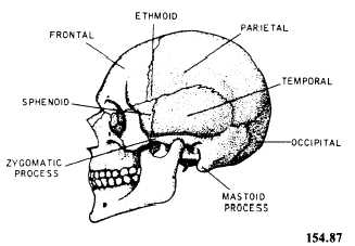
40 lateral view of skull with labels
Northern Lights Holidays - See The Northern Lights In Lapland View our holidays here. Autumn Holidays. Let us help you. For your best chance to see the Northern Lights, contact the UK’s only Aurora specialists. 01670 785012 . or. Email us View all holidays to Finland. Our holidays can be found in numerous locations across Finnish Lapland, all of which have been carefully selected for their excellent Northern Lights potential. Click here to … HUMAN BEING :: ANATOMY :: SKELETON :: LATERAL VIEW OF SKULL image ... lateral view of skull Skull: bony structure enclosing and protecting the brain. The eight cranial bones in an adult are fused to each other by means of sutures. previous next temporal bone Flat skull bone that protects mainly the organs responsible for hearing and equilibrium. frontal bone
Label Parts of the Skull - Lateral View - Printable Label Parts of the Skull - Lateral View - Printable Label Parts of the Skull - Lateral View - Printable Download and print this quiz as a worksheet. You can modify it to fit your needs before you download. Printable Settings Before you print this worksheet you can modify it to your liking using the settings below. Quiz Style Portrait Landscape

Lateral view of skull with labels
Skull Labeling - Answer Key - The Biology Corner Skull Labeling Skull Labeling - Answer Key 1. Coronal Suture 2. Frontal 3. Parietal 4. Nasal 5. Squamosal Suture 6. Ethmoid 7. Lacrimal 8. Sphenoid 9. Lamdoidal Suture 10. Occipital 11. Temporal 12. Zygomatic 13. Maxilla 14. Mandible Solved Identify the bones and features indicated on this - Chegg Identify the bones and features indicated on this lateral view of the skull by clicking and dragging the labels to the correct location. Show transcribed image text Expert Answer Answer : 1. Coronal Suture 2. … View the full answer Solved Label the bones of the lateral view of the skull by | Chegg.com Expert Answer. 96% (24 ratings) Transcribed image text: Label the bones of the lateral view of the skull by clicking and dragging the labels to the correct location. Sphenoid bone Frontal bone Occipital Lacrimal Parietal bone bone Temporal Ethmoid bone MaxillaZygomatic Frontal Sphenoid EthmoidParietal Nasal bone bone bone Maxilla bone bone ...
Lateral view of skull with labels. Lateral Skull Bone Markings Quiz | GetBodySmart Skull lateral markings quiz - Part 1: Lateral Skull Bone Markings Quiz 1. Start Quiz Retake Quiz. Skull lateral markings quiz - Part 2: ... Os Coxa Bone Quiz - Lateral View Markings. Subject Areas. Skeletal System; Muscular System; Nervous System; Urinary System; Circulatory System; Muscle Physiology; Respiratory System; System Quizzes; Surface Enhanced Raman Spectroscopy - an overview S. Uskoković-Marković, ... I. Holclajtner-Antunović, in Encyclopedia of Spectroscopy and Spectrometry (Third Edition), 2017. Abstract. Surface-enhanced Raman scattering (SERS) is achieved when an analyte is adsorbed onto or in close proximity to a prepared roughened metal surface (primarily gold or silver) in a process which greatly enhances the Raman emission. Petrous bone CT: normal anatomy| e-Anatomy - IMAIOS 13.09.2021 · Anatomy of the temporal bone: how to view the anatomical labels. This module is a comprehensive and affordable learning tool for residents and medical students and specially for neuroradiologists and otolaryngologists. It provides images in the axial and coronal planes, allowing the user to review and learn anatomy interactively. Images are ... Right Lateral View of Skull - Quiz - quizizz.com Right Lateral View of Skull. 83% average accuracy. 356 plays. 11th - 12th grade . 3 years ago by . Dominick Damm. 1 Save Share Edit Copy and Edit. QUIZ. NEW. SUPER DRAFT. Right Lateral View of Skull. ... Label letter ----> B. answer choices . parietal bone. squamous suture. lambdoid suture parietal bone
Solved Label the bones and bone features shown on the - Chegg Label the bones and bone features shown on the lateral view of the skull by clicking and dragging the labels to the correct location. Zygomatic process Styloid process External acoustic meatus Temporal bone Lacrimal bone Mandibular condyle Mental foramen Coronoid process Mastoid process Nasal bone Reset Zoom diagram label of skeleton Skull superior diagram cranium labels floor axial skeleton bone blank anatomy atlas human visual labeled physiology bones flickr study. Mandible, lateral view with labels. Blank muscle anatomy physiology diagram label of skeleton. Clinical Practice Guidelines : Child Abuse Diagrams. 11 Pics about Clinical Practice Guidelines : Child Abuse ... Lateral View of Skull - Sinclair Lateral View of Skull. Frontal Bone . Parietal Bone. Ethmoid. Nasal Bone. Lacrimal Bone. Maxilla. Zygomatic Bone. Greater Wing of Sphenoid. Occipital Bone. Mandible. Coronoid Process of Mandible. Condyle of Mandible. Temporal Bone. Mastoid Process of Temporal Bone. External Acoustic Canal (meatus) PREVIOUS (LEFT ARROW) Solved Click and drag the labels to their correct locations | Chegg.com Click and drag the labels to their correct locations on the lateral view of the skull. Styloid process Lacrimal bone Temporal process Mental foramen Mastoid process of temporal bone Mandibular condyle Coronoid process Squamous suture Nasal bone Lambdoid suture External acoustic meatus Coronal suture Reset Zoom
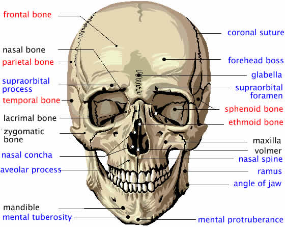
lateral view of skull labeling quiz Diagram | Quizlet lateral view of skull labeling quiz Diagram | Quizlet lateral view of skull labeling quiz STUDY Learn Write Test PLAY Match + − Created by Evan_Finkel2 PLUS Terms in this set (15) nasal bone ... maxilla ... lacrimal bone ... frontal bone ... parietal bone ... incisive bone / premaxilla ... mandible ... tympanic bulla ... occipital bone ... anatomyandphysiologytutor: " A right lateral view of the skull ... Jan 23, 2018 - anatomyandphysiologytutor: " A right lateral view of the skull, complete with labels. Specifically, using this to point out the cranial sutures. Sutures are when two bones have fused together (in... Skull anatomy: Anterior and lateral views of the skull | Kenhub Anterior and lateral views of the skull. The human skull consists of about 22 to 30 single bones which are mostly connected together by ossified joints, so called sutures. The skull is divided into the braincase ( cerebral cranium) and the face ( visceral cranium ). The main task of the skull is the protection of the most important organ in the ... Lateral Skull Bones Quiz | GetBodySmart Lateral Skull Bones Quiz. Start Quiz. Want to speed up your learning and get your anatomy knowledge to the next level? Advanced quizzes are the perfect way to master your exam. Learn anatomy faster and. remember everything you learn. Start Now. <. Quiz - Sacrum and Coccyx Anatomy.

Flickriver
Skull (lateral view) | Radiology Reference Article - Radiopaedia lateral projection centering point the beam travels laterally, with 0° of angulation, through a point ~4 cm above the external auditory meatus collimation superiorly to include skin margins inferiorly to include base of skull anteriorly to include frontal bone posteriorly to the skin margins orientation landscape detector size 24 cm x 30 cm

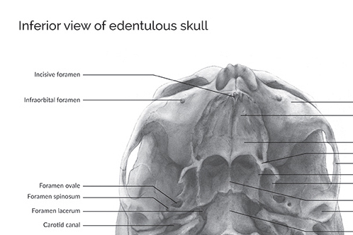
Dentistry lectures for MFDS/MJDF/NBDE/ORE: Diagrams Of Anatomy Of Skull With Radiographic Land Marks
Buy Medical Supplies Medical Equipment -- at Betty Mills Find a large Selection of Medical Supplies, Medical Equipment and Incontinence Aids at Betty Mills. Buy in Bulk and save!
Skull - Anatomy & Physiology
Home Page: The American Journal of Emergency Medicine We use cookies to help provide and enhance our service and tailor content. To update your cookie settings, please visit the Cookie Preference Center for this site.
Divisions Of Skeleton
Solved Identify the bones and features indicated on this - Chegg View the full answer Transcribed image text: Identify the bones and features indicated on this lateral view of the skull by clicking and dragging the labels to the correct location.

Skull, anterior view with labels | A&P | Pinterest | Tags, Axial skeleton and Skulls
Lumbar vertebra, lateral view with labels - Pinterest Apr 1, 2018 - This is Page 55 of a photographic atlas I created as a laboratory study resource for my BIOL 121 Anatomy and Physiology I students on the bones and bony landmarks of the axial skeleton. Credits: All photography, text, and labels by Rob Swatski, Assistant Professor of Biology, Harrisburg Area Community College - York…
Parasellar Skull Base Anatomy | Neuroanatomy | The Neurosurgical Atlas, by Aaron Cohen-Gadol, M.D.
Radiological anatomy of the spine - e-Anatomy - IMAIOS Sep 13, 2021 · Standard radiographic view of anatomical structures of the spinal column. On "Anatomical parts" the user can choose between three types of labels: vertebrae, bones and joints. On "Series" the user can select the radiographs concerning the spine as a whole, the cervical, thoracic and lumbar vertebrae, the sacrum and coccyx.

LATERAL SCAPULA | Radiology, Radiology technologist, Radiography
Infant Skull and Fontanelles - Lateral (Side) View - Innerbody The posterior fontanelle is no longer obvious when the infant is four months old. The anterior fontanelle can normally be felt until 9-16 months of age. Eventually the fontanelles close as the cranial bones grow together. The posterior fontanelle usually closes about two months after birth; the sphenoid fontanelle closes at about three months ...
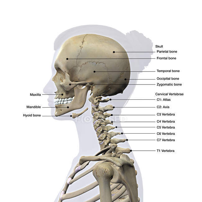
Lateral view of a female skull and cervical spine on white background with labels — illustration ...
Dog Skeletal Anatomy - Sheridan College 04.07.2022 · Acetabular Notch: Notch found on the ventral aspect of the acetabulum.; Acetabulum: A large articulation area with the head of the femur, and divided into Acetabular fossa, Lunatesurface.. Acetabular Fossa: A non-articular depression portion of the acetabulum used for the attachment of the ligament of the head of the femur.; Lunate Surface: Articular …
Wendy Gu Visualizations | Medical and Scientific Illustration and Animation
Gloves at BettyMills Find Gloves On Sale at BettyMills - Get Double Markdown® Savings, Speedy Delivery, plus Valuable Rewards.
Side View of the Skull | ClipArt ETC
Label The Skull (Lateral View) Quiz - PurposeGames.com This is an online quiz called Label The Skull (Lateral View) There is a printable worksheet available for download here so you can take the quiz with pen and paper. From the quiz author. Label all bones, processes, and sutures. Your Skills & Rank. Total Points. 0. Get started! Today's Rank--0.

The Skull - Human Anatomy - GUWS Medical
The Skull Bones - Lateral View | GetBodySmart The Skull Bones - Lateral View Learn anatomy faster and remember everything you learn Start Now Cranial Bones: Frontal bone ( os frontale). Glabella (glabella frontalis) Supercilliary arch or supraorbital ridge (arcus superciliaris frontalis) Zygomatic process (processus zygomaticus frontalis) Lateral view of the frontal bone. 1 2 3 4 5 6 7 8 9

Skull illustration, lateral view - Axial Skeleton Visual A… | Flickr
Lateral view of the brain: Anatomy and functions | Kenhub The lateral view of the brain shows the three major parts of the brain: cerebrum, cerebellum and brainstem.. A lateral view of the cerebrum is the best perspective to appreciate the lobes of the hemispheres. Each hemisphere is conventionally divided into six lobes, but only four of them are visible from this lateral perspective.The lobes are named after the bones of the skull that overlie them:

BRAIN - SAGITTAL SECTION
skull, lateral view, rt side (left label) Diagram | Quizlet Only $2.99/month skull, lateral view, rt side (left label) STUDY Learn Write Test PLAY Match Created by briannaa236 Terms in this set (8) zygomatic process of temporal bone parietal bone temporal bone occipital bone external occipital protuberance mastoid process of temporal bone external acoustic meatus styloid process of temporal bone

Skull Anterior (Markings)
The Adult Skull... Lateral View Quiz - PurposeGames.com This is an online quiz called The Adult Skull... Lateral View There is a printable worksheet available for download here so you can take the quiz with pen and paper. From the quiz author Do you know the different parts of the skull? This quiz has tags. Click on the tags below to find other quizzes on the same subject. Anatomy Bones Skull

Dentistry lectures for MFDS/MJDF/NBDE/ORE: Radiographic Anatomy of Facial Bones and Mandible ...
Anatomical terminology - Wikipedia Anatomical terms used to describe location are based on a body positioned in what is called the standard anatomical position.This position is one in which a person is standing, feet apace, with palms forward and thumbs facing outwards. Just as maps are normally oriented with north at the top, the standard body "map," or anatomical position, is that of the body standing upright, with …

Skull and Bones Medical Illustrations | redbudart
The Skull | Anatomy and Physiology I | | Course Hero The cranium (skull) is the skeletal structure of the head that supports the face and protects the brain. It is subdivided into the facial bones and the brain case, or cranial vault (Figure 1). The facial bones underlie the facial structures, form the nasal cavity, enclose the eyeballs, and support the teeth of the upper and lower jaws.
Post a Comment for "40 lateral view of skull with labels"